.svg)


Introduction
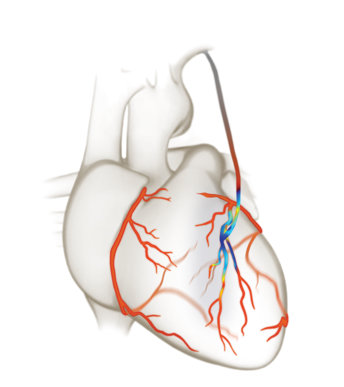
Cardiac surgery today, while advanced, relies on separate tools for visualizing anatomy and measuring blood flow. Correlating information between multiple devices like this leaves room for uncertainty. Envision an innovative instrument seamlessly integrating Transit Time Flow Measurement (TTFM) with high-resolution imaging, revolutionizing cardiac procedures by offering enhanced precision and instantaneous insights. The MiraQ enables surgeons to perform with exceptional accuracy, ultimately leading to improved patient outcomes.
MiraQ is the only instrument combining Transit Time Flow Measurement (TTFM) and high-resolution imaging. It empowers cardiac surgeons with precision and real-time insights, elevating patient outcomes in one innovative solution. Perform cardiac imaging, measure blood flow and receive immediate feedback on how well a graft is functioning.
Intraoperative surgical guidance and quality assessment with ultrasonic imaging and blood flow measurement:
- reduces risk of stroke for the patient.
- provides surgeons with a tool to verify graft functionality, revise when needed and optimize the graft strategy during surgery.
These benefits are displayed in the largest multi-center study to combine TTFM and High Frequency Ultrasound Imaging - the REQUEST Study.
The REQUEST Study
REQUEST is the first multicenter study documenting the changes in surgical strategy from the combined use of Transit Time Flow Measurement (TTFM) and High Frequency Ultrasound Imaging (HFUS).
The results showed that 25% of the patient population had one or more surgical procedure changes made. These changes were associated with low rates of in-hospital mortality and major morbidity. Such an impact on surgical strategy shows the importance of performing routine intraoperative quality assessment with TTFM and HFUS during CABG surgery.

Global approach
Seven leading cardiac surgery centers from Europe, USA and Canada participated. The study was initially led by Professor Pieter Kappetein of Thorax Erasmus MC, Rotterdam, and later by Coordinating Investigator Professor David Taggart from the University of Oxford.
Inclusion criteria was isolated CABG with a minimum of two grafts. Medistim’s VeriQC™ or MiraQ™ devices were used.
7
1016
25%


Download the 4 page Executive Summary of the REQUEST study

Videos

High-Frequency Ultrasound Imaging of aorta prior to manipulation

Graft patency verification with flow measurement and High-Frequency Ultrasound imaging

High-Frequency Ultrasound Imaging of conduits

High-Frequency Ultrasound Imaging of coronary target

Graft patency verification with flow measurement and High Frequency Ultrasound Imaging

High-Frequency Ultrasound Imaging of composite graft

Purse string effect identified with High-Frequency Ultrasound Imaging
Case Studies
Webinars
Publications
Products

MiraQ Cardiac

MiraQ Ultimate
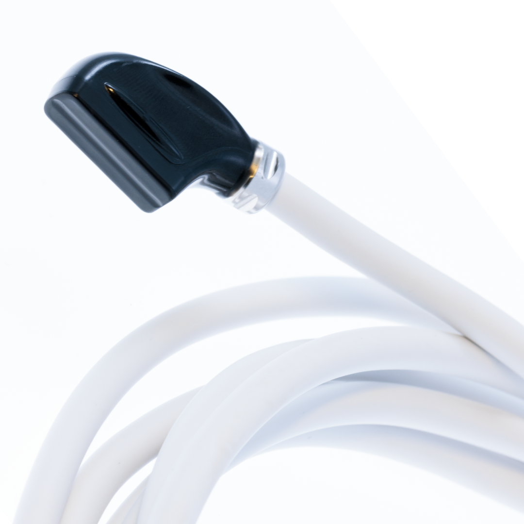
Imaging Probe